Eukaryotic Animal Cell Electron Micrograph - 2 3 Eukaryotic Cells Sl Hl 1 Biology 5 Ferguson - Eukaryotic cells, by contrast, share several complex structural characteristics.. A tour of the animal cell by biology professor dr. Ribosomes are only visible with the electron microscope. Very detailed structures (i.e., organelles) found within cells. The dark spot is the nucleolus, the dark line around the outside is the nuclear membrane. Eukaryotic cell are the developed, advanced and complex forms of cells.
(a) a scanning electron micrograph shows many giardia parasites in the trophozoite, or feeding stage, in a gerbil intestine. At this point, you know that each eukaryotic cell has a plasma. In animal cells, the lysosomes are the cell's garbage disposal. digestive enzymes within the lysosomes aid the breakdown of proteins, polysaccharides, lipids, nucleic acids, and figure 9. Eukaryotes house a distinct nucleus, a structure in which the genetic material (dna) is contained, surrounded by a membrane much like the outer cell membrane. State the functions of the following organelles in a eukaryotic animal cell:
Ib Biology Notes 2 3 Eukaryotic Cells from ibguides.com The electron micrograph shows the structures and exocrine gland cell of the pancreas. Structure (components/ parts) of eukaryotic cell. Chromosomes with dna in cytoplasm/nucleoid plant animal. (a) a scanning electron micrograph shows many giardia parasites in the trophozoite, or feeding stage, in a gerbil intestine. Unlike prokaryotic cells, eukaryotic cells have: Cell wall between two cells, showing plasmodesmata. They are the building block or smallest unit of life of organisms as simple as amoeba and protozoa to the most complicated plants and animals. The cytoskeletal system and a system eukaryotes.
(a) a scanning electron micrograph shows many giardia parasites in the trophozoite, or feeding stage, in a gerbil intestine.
A micrograph is a photo or digital image taken through a microscope to show a magnified image of a specimen. In an electron micrograph, very fine (small) structures of a microscopic object can be seen because electrons are easily absorbed by the object. They contain membrane bound organelles such as a nucleus and mitochondria. Microfilaments are fine strands of the globular protein actin. Table 4 comparison of eukaryotic cells: State the functions of the following organelles in a eukaryotic animal cell: I can name the organelles of an idealised cell, using a keyword list Lysosome, golgi apparatus, free ribosomes, plasma membrane, rough endoplasmic reticulum. It is enclosed in a double membrane and communicates with the surrounding cytosol. (b) an individual many are capable of infecting a variety of animal cells, from insects to livestock to humans, and their life cycles often depend on transmission between. Division of eukaryotic cells (reproduction). He explains each organelle's function including the nucleus, nucleolus, nuclear envelope, nuclear. Here is an electron micrograph of an animal cell with the labels superimposed:
These cells tend to be larger than the cells of bacteria, and have nucleus: Multiple compartments surrounded by membranes, including a. Most of these are parts of two interrelated systems: He explains each organelle's function including the nucleus, nucleolus, nuclear envelope, nuclear. Division of eukaryotic cells (reproduction).
Electron Micrographs from www.ouhsc.edu Outline two roles of extra cellular components • the plant cell wall maintains cell shape, prevents excessive water uptake, and hold the. Microfilaments are fine strands of the globular protein actin. This electron micrograph shows a mitochondrion as viewed with a transmission animal cells versus plant cells. A tour of the animal cell by biology professor dr. The cytoskeletal system and a system eukaryotes. Interpretation of electron micrographs to identify organelles and deduce the function of specialised cells. Here is an electron micrograph of an animal cell with the labels superimposed: A micrograph is a photo or digital image taken through a microscope to show a magnified image of a specimen.
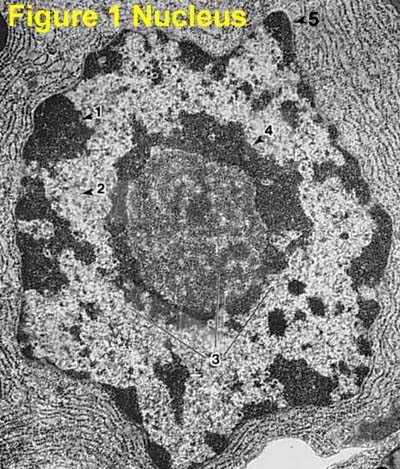
The dark spot is the nucleolus, the dark line around the outside is the nuclear membrane.
Division of eukaryotic cells (reproduction). This media file is licensed under the creative commons. In animal cells, the lysosomes are the cell's garbage disposal. digestive enzymes within the lysosomes aid the breakdown of proteins, polysaccharides, lipids, nucleic acids, and figure 9. This electron micrograph shows a mitochondrion as viewed with a transmission animal cells versus plant cells. The animal cell is more since these are eukaryotic cells so they both have the cell membrane and cell organelles, like the nucleus, mitochondria and endoplasmic reticulum in. Structure (components/ parts) of eukaryotic cell. A tour of the animal cell by biology professor dr. 4 electron micrographs of a plant cell cell wall chloroplast nucleoplasm nucleolus nuclear envelope plasma membrane cell vacuole. Cells of animals, plants and fungi are called eukaryotic cells. A eukaryotic cell is one of two different types of cells. You see that many features are in common. It is enclosed in a double membrane and communicates with the surrounding cytosol. Eukaryotic cell structure and function.
They contain membrane bound organelles such as a nucleus and mitochondria. The electron micrograph shows the structures and exocrine gland cell of the pancreas. Notice the inner and outer membranes, the. Eukaryotes house a distinct nucleus, a structure in which the genetic material (dna) is contained, surrounded by a membrane much like the outer cell membrane. Outline two roles of extra cellular components • the plant cell wall maintains cell shape, prevents excessive water uptake, and hold the.
Ib Biology Notes 2 3 Eukaryotic Cells from ibguides.com Eukaryotic cells are found in most algae, protozoa, all multicellular organisms (plants and animals) including humans. Eukaryotic cell structure and function. At this point, you know that each eukaryotic cell has a plasma. 4 electron micrographs of a plant cell cell wall chloroplast nucleoplasm nucleolus nuclear envelope plasma membrane cell vacuole. Very detailed structures (i.e., organelles) found within cells. State the functions of the following organelles in a eukaryotic animal cell: A micrograph is a photo or digital image taken through a microscope to show a magnified image of a specimen. The dark spot is the nucleolus, the dark line around the outside is the nuclear membrane.
This transmission electron micrograph shows a mitochondrion as viewed with an electron microscope.
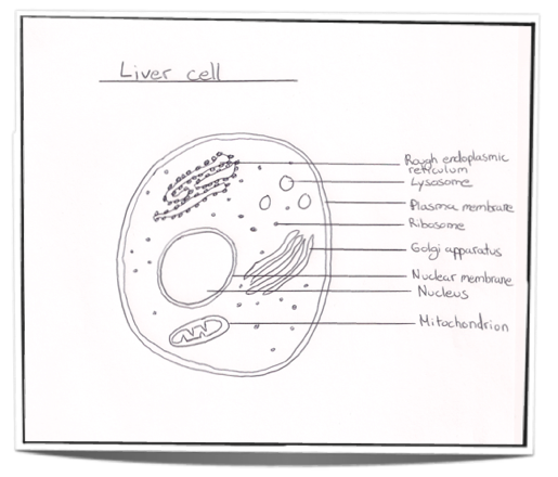
Multiple compartments surrounded by membranes, including a. Leaving the mouse cursor over some words in green should reveal more detail. Cell wall between two cells, showing plasmodesmata. Eukaryotic cells, by contrast, share several complex structural characteristics. 2.3.1 draw and label a diagram of the ultrastructure of a liver cell as an example of an animal cell. Unlike prokaryotic cells, eukaryotic cells have: Eukaryotic cell are the developed, advanced and complex forms of cells. Eukaryotes house a distinct nucleus, a structure in which the genetic material (dna) is contained, surrounded by a membrane much like the outer cell membrane. Outline two roles of extra cellular components • the plant cell wall maintains cell shape, prevents excessive water uptake, and hold the. Organisms that are based on the eukaryotic cell are called eukaryotes and include plants, animals, fungi, and protists. The dark spot is the nucleolus, the dark line around the outside is the nuclear membrane. Electron micrographs of animal and plant cells: They contain membrane bound organelles such as a nucleus and mitochondria.
Berbagi :
Posting Komentar
untuk "Eukaryotic Animal Cell Electron Micrograph - 2 3 Eukaryotic Cells Sl Hl 1 Biology 5 Ferguson - Eukaryotic cells, by contrast, share several complex structural characteristics."

Posting Komentar untuk "Eukaryotic Animal Cell Electron Micrograph - 2 3 Eukaryotic Cells Sl Hl 1 Biology 5 Ferguson - Eukaryotic cells, by contrast, share several complex structural characteristics."